Glaucoma

Man With Glasses Smiling

Glaucoma

Glaucoma is damage to the optic nerve, the cable that transmits sight from the eye to brain, caused by high pressure in the eye. This damage leads to permanent vision loss, typically starting with the peripheral (side) vision then progressing to the central vision.

Normal Eye Compared To An Eye With Glaucoma

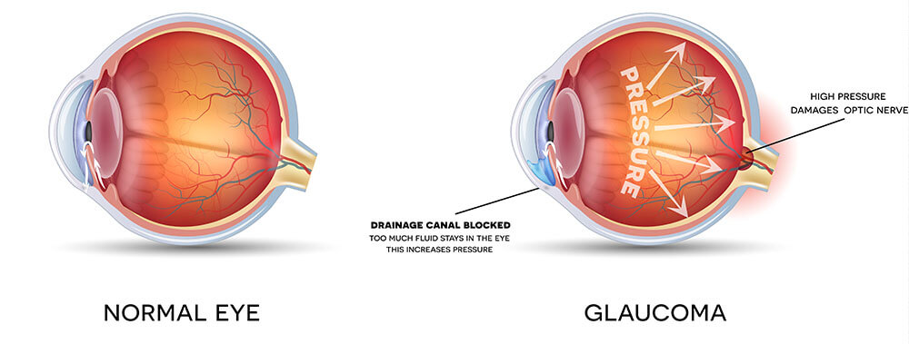
In general, glaucoma results from the eye producing fluid faster than it can drain. The cause depends on the type of glaucoma:

  • Primary Open-Angle Glaucoma (POAG): The most common type of glaucoma, POAG results from the eye’s drainage system clogging over time. Risk factors include age, African-American heritage, and family history. POAG develops slowly and silently; there is no pain, redness, swelling, or other symptoms.
  • Primary Angle-Closure Glaucoma (PACG): Also known as narrow angle glaucoma, PACG results from the iris blocking the drainage system and creating scar tissue. Risk factors include hyperopia (farsightedness) and cataracts. It can develop slowly over time or suddenly with an attack of angle closure that results in severe pain, redness, nausea, headache, and vision loss.
  • Secondary Glaucoma: Results from a secondary cause, such as injury, steroid use, diabetes, or a vessel blockage in the eye.
  • Congenital Glaucoma: Results from abnormal formation of the eye’s drainage system so that it doesn’t work properly. This type of glaucoma is rare.

Most types of glaucoma have no symptoms until significant vision loss has occurred. Therefore, it is critical to get routine eye exams to monitor for development and progression.

Angle closure glaucoma can have a sudden attack that results in severe pain, redness, nausea, headache, and vision loss. If these symptoms develop, you should immediately call your ophthalmologist or go to the nearest emergency room.

There are four ways to detect and monitor glaucoma:

  • Intraocular Pressure: For most individuals, a normal eye pressure is 20 or lower, and anything higher poses a risk for developing glaucoma. Some individuals can develop glaucoma even with a pressure under 20. Your doctor will monitor your pressure to determine your risk for developing glaucoma and your need for additional testing.
  • Optic Nerve Exam: Glaucoma causes damage to the optic nerve, the cable that transmits sight from the eye to the brain. This damage makes the center of the optic nerve look larger, and progressive enlargement can indicate progression of glaucoma. Your doctor will monitor the appearance of the optic nerve and might take photos to compare over time.
  • Optical Coherence Tomography (OCT): This scan measures the thickness of the optic nerve. Glaucoma causes thinning of the nerve, so this scan can monitor for progression.
  • Visual Field Testing: Glaucoma causes permanent vision loss, typically starting with the peripheral (side) vision then progressing to the central vision. Visual field testing monitors loss from glaucoma.

Treatment options depend on the type of glaucoma:

  • Open-Angle Glaucoma: The problem is a clogged drainage system resulting in fluid being produced faster than it is drained. Therefore, the treatment goal is to slow the production of fluid and/or increase the rate of drainage. This can be accomplished through eye drops and/or selective laser trabeculoplasty (SLT), and easy, painless laser procedure done in the office. If these treatments are not sufficient, then next steps include minimally-invasive glaucoma surgery (MIGS) performed during cataract surgery or standalone glaucoma surgery, e.g. trabeculoplasty or tube shunt.
  • Angle-Closure Glaucoma: The problem is the iris blocking the drainage system and creating scar tissue. Therefore, the treatment goal is to move the iris to unblock the drainage system, then to bypass scar tissue if necessary. The iris can be moved with laser peripheral iridotomy (LPI) or cataract surgery. If there is scar tissue and the pressure remains high, then glaucoma surgery might be necessary.

Your doctor will discuss all of the treatment options and help you choose the best options for you.

Four Convenient Locations

South Broad Street

1930 S. Broad St.
Philadelphia, PA 19145

Tel: (215) 339-8100 get directions

Allegheny Ave

2610 E Allegheny Ave
Philadelphia, PA 19134

Tel: (215) 423-5154 get directions

Wills eye Hospital

840 Walnut Street
Philadelphia, PA 19107

Tel: (215) 339-8100 get directions

Wills Northeast Consultation Office

8025 Roosevelt Blvd.
Philadelphia, PA 19152

Tel: (215) 423-5154 get directions

Contact

Get in Touch